New research shows how AI technology can spot breast cancer in MRI scans more accurately than current digital methods, while also pinpointing exactly where suspicious tissue is located — a breakthrough that could make the sensitive screening tool available to more women.
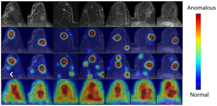
The system takes a novel approach by learning what normal breast tissue looks like, then flagging anything unusual, which is the opposite of how cancer-detection AI has typically been built. When it identifies potential cancer, it creates a visual heatmap showing radiologists precisely where to look.
Researchers with the University of Washington, Microsoft’s AI for Good Lab, and Seattle’s Fred Hutchinson Cancer Center were the lead collaborators on the study. Their results were recently published in Radiology, a journal of the Radiological Society of North America. They trained their AI model on roughly 9,500 MRIs collected at the UW between 2005 and 2022.
The innovation could help expand access to breast MRI screening, which is more sensitive than mammography but currently limited primarily to high-risk patients due to cost and efficiency concerns.
“We’re hoping to be able to offer breast MRI to more women than we do nowadays because it is a really sensitive breast screening tool,” said Savannah Partridge, a UW professor of radiology. “But to do that, we’re looking at how do we scale?”
The strategy for building the model flipped the traditional approach on its head. Instead of learning to detect scans that are positive for cancer, the model was trained to recognize normal, or benign, images and then flag MRIs that included abnormal cells.
The approach, called “anomaly detection,” makes sense given that researchers have many more non-cancerous images than those showing disease, said Partridge, “so we’re able to leverage our data more efficiently.”
An essential feature of the new tool is that it creates a heatmap overlaying the image, visually highlighting the area of concern. Other technologies sometimes indicate only whether cancer was detected in an MRI, but not precisely where it was found.
“Our model provides an understandable, pixel-level explanation of what’s abnormal in a breast,” said Felipe Oviedo, a senior research analyst at Microsoft’s AI for Good Lab, in a statement.
The AI analysis could help radiologists prioritize cases that need quicker attention, guide providers in additional imaging, or indicate an area that requires a biopsy.
The tool is not ready for use in clinical settings. Researchers are planning additional studies to see how the technology performs against radiologists reviewing the same images to better understand its benefits.
Partridge, who is the UW’s research director of breast imaging and the past associate director of cancer imaging at Fred Hutch, said the collaboration with Microsoft gave her the chance to be closely involved with the creation of the algorithm, providing insight into how it was built and behaved.
Still, Partridge wants to proceed with caution when adopting AI for healthcare, ensuring that any clinical tool provides trustworthy, useful information that supports radiologists, rather than complicating their work.
“It’s not do you use [AI], or do you not, but how do you use it?” she said. “How do you use it appropriately and safely?”
Additional authors of the study, which was titled “Cancer Detection in Breast MRI Screening via Explainable AI Anomaly Detection,” are Anum Kazerouni, Philipp Liznerski, Yixi Xu, Michael Hirano, Robert Vandermeulen, Marius Kloft, Elyse Blum, Adam Alessio, Christopher Li, William Weeks, Rahul Dodhia, Juan Lavista Ferres and Habib Rahbar. Their affiliations include the UW, Microsoft, Fred Hutch, Michigan State University, University of Kaiserslautern-Landau, Berlin Institute for the Foundations of Learning and Data, and Technical University of Berlin.
Read the full article here